Yazılar

Scientists Develop Advanced 3D Maps to Explore Octopus Arm Mechanics

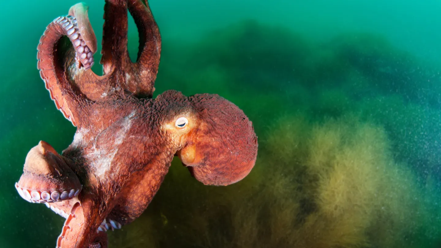
Researchers at San Francisco State University have made groundbreaking advancements in our understanding of octopus arm mechanics by developing intricate three-dimensional maps that illustrate the complex nervous system within these remarkable appendages. Unlike human limbs, which are entirely controlled by the brain, octopus arms exhibit a high degree of autonomy, enabling them to perform intricate tasks with limited direct input from the central nervous system. This semi-independent functionality allows octopuses to execute actions such as opening jars and manipulating tools, showcasing their remarkable adaptability in diverse environments.

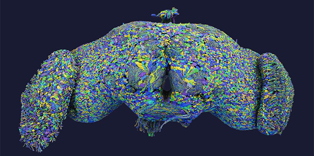
The study, led by Robyn Crook, Associate Professor and Associate Chair of the SF State Biology Department, addresses a critical question in marine biology: how do octopus arms manage to perform such complex behaviors without constant communication with the brain? To uncover the secrets of this neural independence, researchers employed advanced 3D imaging techniques. Gabrielle Winters-Bostwick, a postdoctoral fellow, and Diana Neacsu, a graduate student, collaborated to create comprehensive anatomical and molecular maps, revealing the distinctive organization of octopus arms.

Winters-Bostwick’s research focused on the functional differentiation of neurons within the arm. By using molecular tags to highlight various types of neurons, she discovered that the neurons located at the tip of the arm are fundamentally different from those situated near the central brain. This finding suggests a sophisticated level of specialization that enables the arm to react to stimuli and perform tasks autonomously. Meanwhile, Neacsu utilized 3D electron microscopy to delve deeper into the structural organization of the arm, identifying repeating patterns in nerve branches and ganglia. These patterns indicate a complex network that may facilitate the arm’s independent operations.

The implications of this research extend beyond the realm of octopuses, offering valuable insights into the evolution of neural control in cephalopods and other organisms. By understanding how octopus arms function with such autonomy, scientists can gain a better appreciation of the evolution of motor control and the potential for similar mechanisms in other species. As researchers continue to explore the depths of octopus biology, the innovative mapping techniques developed in this study could pave the way for future investigations into the nervous systems of other complex organisms, enhancing our knowledge of the diverse strategies life employs to thrive in various environments.

Research Uncovers How the Brain Segments Continuous Experiences into ‘Scenes’

New Study Reveals How the Brain Segments Experiences into Distinct Moments

Recent research has shed light on the fascinating mechanisms by which our brains organize daily experiences into meaningful segments, akin to scenes in a movie. While we often perceive life as a seamless flow of events, this study reveals that our brains automatically parse these experiences into distinct moments. The longstanding debate among scientists has revolved around whether these memory boundaries are dictated by changes in the environment or shaped by personal interpretation. However, a study led by Christopher Baldassano, an associate professor of psychology at Columbia University, provides compelling evidence that our brains actively select these transitions based on our goals and prior experiences.

To delve deeper into how the brain delineates memories, Baldassano and his research team employed functional magnetic resonance imaging (fMRI) in a carefully designed experiment. Participants were asked to listen to various narratives that portrayed key social scenarios, including a business deal, a marriage proposal, and a breakup, while their brain activity was closely monitored. The focus of the research was primarily on the medial prefrontal cortex (mPFC), a region known to play a critical role in processing ongoing events and understanding social contexts.

The findings from this study were revealing. The data indicated that significant social events within the narratives, such as the successful conclusion of a business deal, were associated with spikes in brain activity. These spikes signified a mental shift, suggesting that the brain recognized a transition point in the story. Furthermore, when participants were prompted to concentrate on specific details, such as the locations mentioned in the narratives, their brain activity shifted accordingly. This demonstrated that the way we segment our experiences is not only influenced by the events themselves but also by our attentional focus and the objectives we set while engaging with the material.

This research holds profound implications for understanding memory formation and retrieval. It suggests that our cognitive processing of experiences is more dynamic and subjective than previously thought, shaped by both external stimuli and internal goals. The insights gained from this study could pave the way for developing strategies to enhance memory retention and recall, particularly in educational settings. As we continue to unravel the complexities of how the brain organizes our experiences, this research highlights the importance of both context and personal interpretation in shaping our memories and understanding of the world.

Groundbreaking Brain Map Unveils 140,000 Neurons and New Nerve Cell Types in Fruit Fly Brain

Researchers Unveil Most Comprehensive Brain Map of Fruit Fly, Identifying 140,000 Neurons and New Nerve Cell Types

Scientists have achieved a remarkable milestone in neuroscience by creating the most detailed map of a fruit fly’s brain ever produced. This intricate map, which reveals nearly 140,000 neurons and an astounding 54.5 million synapses, is the culmination of over four years of dedicated research led by neuroscientists Mala Murthy and Sebastian Seung at Princeton University. Known as a ‘connectome,’ this map stands as the most complete brain diagram for any organism to date, offering unprecedented insights into the complexity of neural networks.

The research team utilized advanced electron microscopy to capture images of the fly’s brain, allowing them to reconstruct its intricate structure. To manage the vast amount of data, AI-assisted tools were employed, although significant portions of the map required meticulous manual editing. In total, the team and their volunteers conducted more than three million manual adjustments to ensure the map’s accuracy and reliability. This rigorous process led to the identification of 8,453 distinct neuron types, of which 4,581 were previously unknown, expanding our understanding of fruit fly neurobiology.

One of the most intriguing findings from this study was the unexpected interconnectivity of various neurons. Researchers discovered that neurons typically associated with specific sensory functions, such as vision, frequently formed connections with neurons that process other sensory inputs like hearing and touch. This interconnectedness suggests a sophisticated integration of sensory information, emphasizing the brain’s ability to process and respond to a multi-faceted environment, which is crucial for the survival of the fruit fly.

The insights gained from the connectome have already proven valuable in simulating fruit fly behavior within virtual models. In a groundbreaking experiment, the simulations demonstrated how neurons involved in taste perception—specifically those detecting sweet and bitter stimuli—activate the motor neurons that control the fly’s proboscis. Remarkably, when compared to actual fly behavior, the virtual model achieved over 90% accuracy in predicting neuronal responses and subsequent actions. This advancement not only enhances our understanding of fruit fly behavior but also paves the way for future research into the neural underpinnings of more complex organisms, including humans.