3D Scanning Technology Uncovers New Details of Endurance Shipwreck After 107 Years

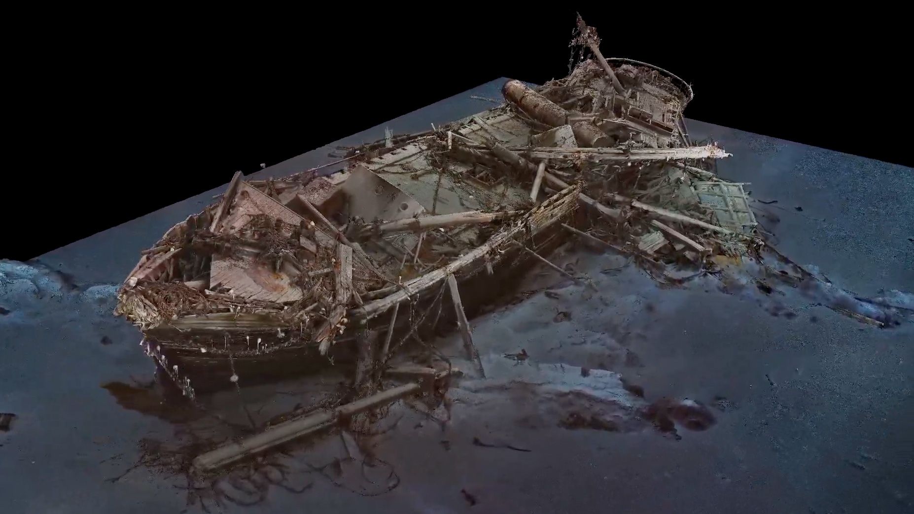
The wreck of the Endurance, famously lost during Sir Ernest Shackleton’s ill-fated Antarctic expedition in 1914, has been meticulously documented using cutting-edge 3D scanning and underwater photography techniques. The ship, which rested on the floor of the Weddell Sea for over a century, was rediscovered in 2022, reigniting interest in its storied history. Now, thanks to the Falklands Maritime Heritage Trust, detailed scans reveal astonishingly well-preserved elements of the 144-foot vessel, including sections of its upper deck that appear remarkably intact despite being submerged for 107 years.

The 3D imaging provides a unique view of the shipwreck as it rests at the bottom of the sea. While some parts, such as the mast and railings, show signs of decay, many artifacts remain eerily well-preserved. Items like scattered plates on the deck and a boot entangled in the ship’s collapsed rigging paint a vivid picture of the ship’s final moments. Notably, fragments of the ship’s linoleum floor, featuring a star-pattern design, are still discernible, highlighting the craftsmanship of the time. These scans are part of a documentary premiering on November 1, which chronicles the shipwreck’s discovery and the gripping survival story of its crew.

Shackleton’s Antarctic expedition aimed to make a historic crossing of Antarctica on foot. However, fate intervened when the Endurance became trapped in dense sea ice before reaching the continent. After enduring ten harrowing months trapped in the ice, the ship was eventually crushed and sank, leaving Shackleton and his crew stranded in one of the harshest environments on Earth. With limited supplies and hope dwindling, Shackleton, alongside five crew members, undertook a perilous journey of over 800 miles in a small lifeboat to reach South Georgia Island, where they sought help. Remarkably, all crew members ultimately survived this extraordinary tale of endurance and leadership.

The preservation of the Endurance serves as a testament to the resilience of both the ship and its crew. As researchers continue to analyze the wreck, the findings not only deepen our understanding of maritime history but also shed light on the challenges faced by early explorers in uncharted territories. The images and data captured through advanced scanning technology offer a window into the past, allowing us to appreciate the rich legacy of exploration and the human spirit’s unyielding determination in the face of adversity.

New Research Discovers Essential Role of Selfish DNA (LINE-1) in Early Human Embryonic Development

A recent study has significantly altered our understanding of transposable elements in the human genome, revealing their vital role in early embryonic development. Researchers at Sinai Health have discovered that these segments of DNA, commonly referred to as “selfish DNA,” particularly the LINE-1 (Long Interspersed Nuclear Element-1), are not merely parasitic but essential for the proper formation of human embryos. Historically viewed as detrimental, these transposable elements constitute approximately 20% of the human genome, while functional genes represent less than 2%, indicating a much more complex relationship than previously thought.

Transposons, the genetic elements capable of relocating within the genome, were once compared to viruses due to their ability to replicate and potentially disrupt normal gene function. However, Dr. Juan Zhang, a senior co-author and postdoctoral fellow involved in the study, pointed out that LINE-1 RNA shows significant activity during the early stages of embryonic development. This observation challenges the long-held belief that transposable elements primarily serve as harmful agents contributing to diseases such as cancer and hemophilia.

The study’s findings highlight the importance of LINE-1 in embryo development, particularly through experiments that involved blocking its activity. When Dr. Zhang’s team inhibited LINE-1 in human embryonic stem cells, they observed that the cells reverted to an earlier developmental stage known as the 8-cell stage. At this stage, the cells are capable of developing into both embryonic and placental tissues, underscoring the necessity of LINE-1 in guiding the differentiation process of cells into specialized forms required for further embryonic development.

These insights shed light on the previously underestimated roles of transposable elements, suggesting that what was once considered “junk DNA” may actually be crucial to the complexities of human development. The implications of this research extend beyond embryology; they may also provide new avenues for understanding genetic diseases and advancing regenerative medicine. By unraveling the functions of LINE-1 and similar elements, scientists can better comprehend their contributions to both normal development and pathological conditions, ultimately leading to innovative therapeutic strategies.

Scientists Develop Advanced 3D Maps to Explore Octopus Arm Mechanics

Researchers at San Francisco State University have made groundbreaking advancements in our understanding of octopus arm mechanics by developing intricate three-dimensional maps that illustrate the complex nervous system within these remarkable appendages. Unlike human limbs, which are entirely controlled by the brain, octopus arms exhibit a high degree of autonomy, enabling them to perform intricate tasks with limited direct input from the central nervous system. This semi-independent functionality allows octopuses to execute actions such as opening jars and manipulating tools, showcasing their remarkable adaptability in diverse environments.

The study, led by Robyn Crook, Associate Professor and Associate Chair of the SF State Biology Department, addresses a critical question in marine biology: how do octopus arms manage to perform such complex behaviors without constant communication with the brain? To uncover the secrets of this neural independence, researchers employed advanced 3D imaging techniques. Gabrielle Winters-Bostwick, a postdoctoral fellow, and Diana Neacsu, a graduate student, collaborated to create comprehensive anatomical and molecular maps, revealing the distinctive organization of octopus arms.

Winters-Bostwick’s research focused on the functional differentiation of neurons within the arm. By using molecular tags to highlight various types of neurons, she discovered that the neurons located at the tip of the arm are fundamentally different from those situated near the central brain. This finding suggests a sophisticated level of specialization that enables the arm to react to stimuli and perform tasks autonomously. Meanwhile, Neacsu utilized 3D electron microscopy to delve deeper into the structural organization of the arm, identifying repeating patterns in nerve branches and ganglia. These patterns indicate a complex network that may facilitate the arm’s independent operations.

The implications of this research extend beyond the realm of octopuses, offering valuable insights into the evolution of neural control in cephalopods and other organisms. By understanding how octopus arms function with such autonomy, scientists can gain a better appreciation of the evolution of motor control and the potential for similar mechanisms in other species. As researchers continue to explore the depths of octopus biology, the innovative mapping techniques developed in this study could pave the way for future investigations into the nervous systems of other complex organisms, enhancing our knowledge of the diverse strategies life employs to thrive in various environments.